
Why Spinal Instability Gets Missed Without Proper Imaging
A patient can leave an emergency department or medical office after a car accident with reassuring news: “Nothing is broken.” Yet days or weeks later, neck pain remains. Headaches continue. The back feels weak or unreliable. In some patients, spinal instability after a car accident may be one of the injuries that has not yet been adequately evaluated.
Sometimes the problem is not that the initial evaluation was wrong. It is that the evaluation answered a different question.
A routine X-ray can be excellent for identifying fractures, alignment abnormalities, and other structural findings. An MRI can provide important information about discs, nerves, the spinal canal, soft tissues, and other anatomy. But spinal injuries are not always fully explained by what the spine looks like while the patient is lying or standing still.
In some trauma patients, an important question is:
What happens when the injured spinal segments move?
That distinction matters because spinal instability can involve abnormal or excessive motion between vertebrae following injury to the structures responsible for controlling that motion. When the imaging strategy does not evaluate the suspected injury appropriately, clinically important findings can be overlooked.
What Spinal Instability Really Means
The spine is designed to move. Flexion, extension, rotation, and side bending are normal and necessary.
But that movement must remain controlled.
Ligaments, joint capsules, muscles, discs, and the geometry of the spinal joints work together to maintain stability while allowing normal motion. Trauma can damage components of this stabilizing system, particularly the ligamentous and capsular structures that help restrain excessive movement.
The distinction is important:
Normal mobility is controlled movement. Instability is loss of adequate motion control.
A patient can therefore have substantial range of motion and still have abnormal segmental mechanics. Conversely, restricted overall range of motion does not rule out abnormal motion at an individual spinal segment.
This is one reason spinal instability cannot be determined simply by asking whether the patient can turn or bend the neck.
Why Car Accidents Can Produce Ligament Injury
Motor vehicle collisions expose the cervical and lumbar spine to rapidly changing forces.
During a collision, different regions of the body accelerate and decelerate at different times. The resulting forces can produce complex combinations of flexion, extension, translation, compression, and rotation.
Ligaments play an important role in limiting excessive vertebral motion. When sufficient force is applied, those tissues can be sprained, stretched, or disrupted.
The patient does not need to sustain a fracture for a meaningful soft-tissue injury to occur.
This is an important distinction because the absence of fracture answers one question:
Is a fracture visible?
It does not automatically answer another:
Are the structures controlling spinal motion functioning normally?
Why Routine Imaging May Not Tell the Whole Story
Every imaging study has a purpose.
Neutral radiographs provide valuable information about alignment, fracture, degeneration, and spinal structure. MRI provides detailed information about discs, neural structures, the spinal canal, bone marrow, and many soft tissues.
But these studies are generally obtained in a relatively static position.
If the clinical concern involves abnormal motion, a static image may not fully evaluate that question.
This does not mean that every patient involved in a car accident requires specialized or dynamic imaging. Imaging should be selected according to the history, mechanism of injury, examination findings, symptoms, prior studies, and clinical indications.
It does mean that persistent symptoms should not automatically be dismissed simply because an initial study did not demonstrate a fracture or another obvious structural lesion.
Sometimes the next question needs a different diagnostic tool.
The Role of Flexion and Extension Imaging
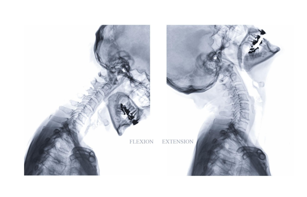
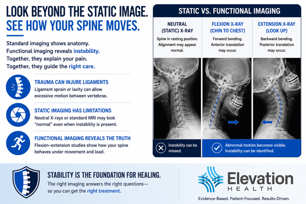
Flexion and extension radiographs allow clinicians to evaluate the relationship between vertebrae as the spine moves through controlled positions.
When clinically indicated, flexion-extension X-rays for spinal instability can provide information about abnormal motion that cannot be obtained from a neutral radiograph alone.
The clinician can for cervical instability by evaluating segmental translation, angular motion, alignment changes, and other biomechanical relationships. Findings must then be interpreted in the context of the patient’s examination, symptoms, trauma history, and other diagnostic studies.
This is particularly important after trauma because the issue may not simply be where a vertebra is positioned at rest.
The issue may be how that vertebra behaves during movement.
MRI Answers a Different Set of Questions
MRI and motion-based radiographic studies should not be viewed as competing tests. They provide different types of information.
MRI may demonstrate disc protrusions or herniations, annular injury, stenosis, nerve involvement, bone marrow changes, and selected ligamentous or soft-tissue abnormalities.
When clinically warranted, specialized MRI protocols or positional studies may provide additional information that routine protocols do not capture.
The goal is not to order more imaging.
The goal is to order the appropriate imaging for the clinical question being investigated.
That is an important difference.
Upper Cervical Injuries Require Particular Attention
The upper cervical region—the occiput, atlas (C1), and axis (C2)—is biomechanically different from the lower cervical spine.
This region allows substantial head and neck motion and depends heavily upon ligamentous structures for stability.
Following trauma, patients with upper cervical involvement may report symptoms including persistent upper-neck pain, headaches, dizziness, balance disturbance, or a sense of cervical fatigue or instability. These symptoms are not specific to ligament injury and may have several possible causes, so they require appropriate clinical evaluation.
When the history and examination raise concern for injury in this region, the imaging strategy should be selected specifically to answer that question.
A routine cervical study should not automatically be assumed to answer every upper-cervical biomechanical question.
The Clinical Examination Still Matters
Imaging should never replace a comprehensive examination.
Range of motion, pain response, muscle strength, neurological findings, balance, functional limitations, and other objective findings help establish the clinical context in which imaging is interpreted.
This becomes especially important in patients whose symptoms persist.
For example, measurable reductions in range of motion, altered pain thresholds, muscle weakness, impaired balance, or loss of functional capacity may demonstrate that a patient’s problem extends beyond a subjective complaint of pain.
No single test tells the entire story.
The strongest clinical picture emerges when the history, examination, functional testing, and imaging findings are evaluated together.
Why Missing Instability Can Change Treatment
A diagnosis should influence treatment.
If an injured segment is suspected or demonstrated to have abnormal motion, treatment planning should account for that finding.
The clinical objective may shift toward protecting injured tissues, improving muscular control and stabilization, correcting relevant biomechanical dysfunction, and progressively restoring activity tolerance rather than simply attempting to increase motion.
This is one reason identifying the underlying injury matters.
Two patients can both say, “My neck hurts,” while having very different injuries requiring different management strategies.
Treating the symptom without identifying the underlying problem can lead to repeated episodes of temporary improvement without resolving the factors contributing to persistent dysfunction.
Persistent Pain Deserves an Explanation
One of the most frustrating experiences after a car accident is being told that everything looks normal while knowing that normal activities still produce symptoms.
Persistent pain does not prove spinal instability. Neither does a normal initial study prove that instability exists.
Both conclusions would go too far.
Instead, persistent or worsening symptoms should prompt a more fundamental question:
Does the available evidence adequately explain what this patient is experiencing?
If the answer is no, further clinical evaluation may be appropriate.
That could mean reviewing the original imaging, performing additional objective testing, obtaining a specialist consultation, or selecting another imaging study based upon the unresolved clinical question.
The Elevation Health Approach
At Elevation Health, the objective is not to order every available test. It is to determine what information is necessary to understand the injury.
For patients recovering from motor vehicle collisions, that means correlating the mechanism of injury with the patient’s symptoms, examination findings, functional deficits, and available imaging.
When those pieces do not adequately explain the patient’s condition, additional evaluation may be considered.
This approach is especially important when symptoms persist despite treatment or when the patient’s functional limitations appear greater than the findings documented during the initial evaluation.
The goal is not more testing. The goal is better answers.
Seeing What a Static Picture Cannot Always Show
A photograph can tell you where something is.
It cannot always tell you how it moves.
That is the central problem when evaluating a potentially dynamic spinal injury solely from static information.
Neutral X-rays, MRI, functional studies, physical examination, and the patient’s history each provide a different piece of the clinical picture. The value comes from determining which pieces are necessary—and then connecting them.
For patients injured in car accidents, this can mean the difference between repeatedly treating unexplained symptoms and finally understanding why those symptoms persist.
Stability Is Part of the Foundation for Recovery
Successful recovery begins with understanding what was injured.
When spinal instability after a car accident is clinically suspected, the evaluation should be designed to determine whether abnormal segmental motion or ligamentous injury is contributing to the patient’s condition.
That does not begin with assumptions.
It begins with the right questions, objective examination, and—when clinically indicated—the right imaging.
Because before you can develop the right treatment plan, you have to understand the injury you are treating.
References
- White AA III, Panjabi MM. Biomechanical Analysis of Clinical Stability in the Cervical Spine. Spine (Phila Pa 1976). 1978;3(2):76–86.
- AAPM&R Now. Spinal Instability Definition, Theory, and Assessment of Spinal Column Function and Dysfunction. 2024.
- Dvorak J, Froehlich D, Penning L, et al. Functional radiographic diagnosis of the cervical spine: flexion/extension. Spine. 1988;13:748–755.