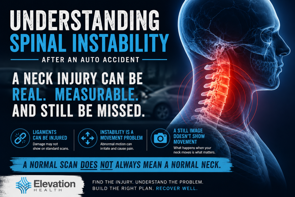
Understanding Spinal Instability After an Auto Accident
Why some neck injuries remain hidden—and why movement matters.
Most people assume that if an X-ray, CT scan, or MRI is normal, their neck is fine. Unfortunately, that’s not always true.
After an auto accident, ligaments that stabilize the cervical spine (neck) can be stretched or torn, causing cervical instability. These injuries may not appear on standard imaging because those tests capture the spine while it is still—not while it is moving.
When injured ligaments allow excessive motion between the vertebrae, the condition is called spinal instability. For some patients, this can become an overlooked source of ongoing neck pain, headaches, dizziness, and other symptoms.
Understanding how spinal instability develops is the first step toward receiving the right diagnosis and the right treatment.
Figure 1. Spinal instability is a movement problem. A neck may appear normal on a static scan while abnormal motion occurs because of ligament injury. Dynamic imaging may reveal instability that static imaging cannot.
What Keeps Your Neck Stable?
Your neck is made up of seven bones called vertebrae. While these bones provide structure, they rely on a network of ligaments, muscles, and joints to keep each vertebra moving in a controlled and coordinated way.
Ligaments are especially important because they act like strong bands that limit excessive movement. During a rear-end or side-impact collision, the head and neck can accelerate and decelerate so quickly that these ligaments may stretch beyond their normal limits or tear, leading to neck instability after whiplash.
When this happens, the spine may no longer move the way it was designed to. Even small changes in movement can place additional stress on muscles, joints, discs, and nerves, contributing to ongoing pain and dysfunction.
Why a Normal Scan Can Miss a Real Injury
Many people are told their X-rays, CT scans, or MRI are normal after an auto accident. While these tests are excellent for detecting fractures, severe disc injuries, and other structural damage, they are not designed to measure how the spine moves.
Spinal instability is a movement problem. When the ligaments that stabilize the neck are stretched or torn, excessive motion can occur between the vertebrae. That abnormal movement may not be visible on routine imaging performed while the neck is at rest, and may not lead to an accurate diagnosis after an auto accident.
Figure 1 compares a stable cervical spine with one affected by spinal instability. The difference is not simply what the spine looks like—it’s how it functions during movement.
![Infographic comparing stable cervical [neck] motion with spinal instability after an auto accident, showing how ligament injury can cause abnormal movement.](https://elevationhealthclinic.com/wp-content/uploads/2026/07/spinal-instability-vs-stable-neck-motion.webp-1024x683.png)
Figure 2. Spinal instability is a movement problem. A neck may appear normal on a static scan while demonstrating abnormal motion due to ligament injury during movement.
Understanding Spinal Instability After an Auto Accident: Why Movement Matters
Clinical Insight
Spinal instability is best evaluated with dynamic imaging. While standard MRI and CT scans remain essential for identifying structural injuries, dynamic flexion-extension X-rays can reveal abnormal motion that static imaging cannot. When clinically indicated, specialized MRI studies may provide additional information about ligament integrity and associated soft tissue injuries.
Diagnosing spinal instability begins with a thorough history and physical examination, but confirming the diagnosis often requires imaging that evaluates motion, not just anatomy.
Standard X-rays, CT scans, and MRI studies are excellent for identifying fractures, significant disc injuries, arthritis, and other structural abnormalities. However, these tests are usually performed while the patient is lying still or standing in a neutral position. Because spinal instability is a movement disorder, static imaging may appear normal even when excessive motion exists between the vertebrae.
When ligament injury is suspected, dynamic flexion-extension X-rays (forward and backward bending of the neck) are commonly used to evaluate abnormal movement between the cervical vertebrae. These studies can demonstrate excessive translation or angulation that may indicate spinal instability.
In selected cases, specialized MRI studies may provide additional information about ligament injury or associated soft tissue damage. These findings are interpreted together with the patient’s history, physical examination, and other imaging studies to determine whether spinal instability is present.
The diagnosis should never rely on a single test. It requires correlating the patient’s symptoms with objective clinical findings and appropriate imaging to determine whether abnormal motion is contributing to their condition.
Key Takeaways
- Spinal instability is a movement problem. Injured ligaments can allow excessive motion between the vertebrae, even when routine imaging appears normal.
- A normal MRI or CT scan does not always rule out ligament injury. Standard imaging is performed while the neck is still and may not reveal abnormal motion.
- Dynamic flexion-extension X-rays can identify spinal instability. When ligament injury is suspected, these studies may reveal abnormal motion that static imaging cannot.
- An accurate diagnosis requires more than one test. Your history, physical examination, and appropriate imaging should be interpreted together.
- Early recognition matters. Identifying spinal instability early helps guide appropriate treatment and may improve long-term outcomes.
References
- Tominaga Y, et al. (2006)
Neck Ligament Strength Is Decreased Following Whiplash Trauma.- Supports that whiplash reduces cervical ligament strength.
- Ivancic PC, et al. (2008)
Whiplash Causes Increased Laxity of the Cervical Capsular Ligament.- Demonstrates increased ligament laxity after whiplash.
- Stemper BD, et al. (2006)
Anterior Longitudinal Ligament Injuries in Whiplash May Lead to Cervical Instability.- Supports the mechanism by which ligament injury may contribute to cervical instability.