
Understanding Ligament Injuries and the Need for Specialized MRI
When patients continue to experience neck pain, headaches, dizziness, or instability after being injured in a car accident, the problem is often deeper than muscle strain or disc irritation. In many cases, the missing piece is ligament injury—and standard imaging is not always sufficient to identify it.
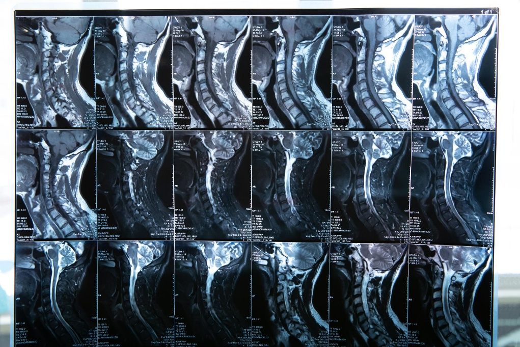
This is where specialized MRI techniques, such as flexion/extension imaging and upper cervical proton density (PD) sequences, become critically important.
Why Ligament Injuries Are Commonly Missed
Ligaments are the stabilizing structures of the spine. They limit excessive motion and protect joints, discs, and the nervous system. During a car accident, especially whiplash-type events, ligaments are often subjected to forces beyond their normal tolerance.
Unlike muscles, ligaments:
- Heal slowly
- Do not regenerate well
- Can remain permanently lax if injured
The challenge is that ligament injuries do not always appear clearly on standard MRI studies. Many conventional MRIs are performed with the patient lying flat, in a neutral position, and using sequences optimized for discs—not stability.
As a result, patients may be told their MRI is “normal” despite ongoing symptoms.
Why Standard MRI Has Limitations
Traditional MRI is excellent for identifying:
- Disc herniations
- Nerve compression
- Major structural abnormalities
However, it has limitations when evaluating functional instability and subtle ligament damage. This is because:
- The spine is imaged in a static position
- Abnormal motion is not assessed
- Certain ligaments are difficult to visualize without specific sequences
For patients with post-traumatic instability, the problem may only become apparent when the spine is moving or loaded, not when it is lying still.
The Role of Flexion/Extension MRI
Flexion/extension MRI evaluates the spine in different positions—typically bending forward (flexion) and backward (extension). This allows doctors to assess how spinal segments behave under movement.
Flexion/extension MRI can reveal:
- Abnormal joint translation
- Excessive motion between vertebrae
- Segmental instability that is not visible in neutral imaging
- Dynamic stress on the spinal cord or nerve roots
For patients injured in car accidents, this type of imaging is especially valuable because ligament injuries often allow too much motion, not too little.
If instability is present, treating the spine as if it were simply “stiff” can worsen symptoms rather than improve them.
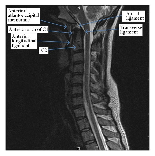
Upper Cervical Injuries Require Specialized Imaging
The upper cervical spine—particularly the relationship between the skull, C1, and C2—is highly specialized and uniquely vulnerable to trauma. These segments rely heavily on ligaments for stability and contain dense neurological structures.
Standard cervical MRI protocols may not adequately visualize:
- Alar ligaments
- Transverse ligament
- Capsular ligaments of the upper cervical joints
This is where proton density (PD) MRI sequences become important.
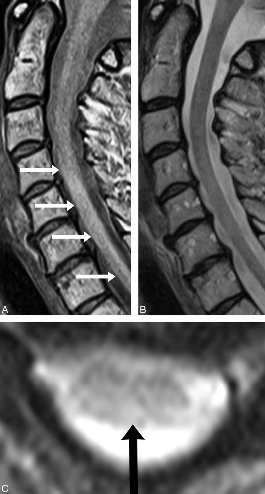
Why Proton Density (PD) MRI Matters
Proton density MRI is optimized to highlight soft tissue contrast, making it particularly useful for evaluating ligaments and joint capsules.
Upper cervical PD imaging can help identify:
- Ligament thickening or disruption
- Signal changes suggesting sprain or tearing
- Asymmetry between stabilizing structures
- Inflammatory changes related to trauma
For patients with persistent headaches, dizziness, neck instability, or neurological symptoms after a crash, these findings can be clinically significant.
Ligament Injury and Spinal Instability
When ligaments are injured, the spine may become unstable. This instability places abnormal stress on:
- Discs
- Facet joints
- Muscles
- The spinal cord and nerve roots
Over time, this can lead to:
- Chronic pain
- Recurrent flare-ups
- Early degenerative changes
- Reduced tolerance to daily activities
Identifying ligament injury early allows care to shift toward stabilization and structural correction, rather than aggressive mobilization.
Why Specialized MRI Changes Treatment Strategy
Knowing whether ligament injury or instability exists fundamentally changes how care should be delivered. For example:
- Excessive manipulation may be avoided
- Stabilization and corrective strategies are prioritized
- Rehabilitation is progressed more cautiously
- Long-term correction becomes the focus, not short-term relief
Without this information, treatment may be incomplete—or even counterproductive.
The Elevation Health Perspective
At Elevation Health, imaging decisions are driven by clinical findings and patient history, particularly in motor vehicle collision cases.
When symptoms persist or instability is suspected, specialized MRI options—such as flexion/extension studies and upper cervical proton density imaging—may be recommended to ensure that ligament injuries are not overlooked.
Patients are educated on:
- Why standard imaging may be insufficient
- What specialized MRI evaluates
- How findings influence corrective care decisions
Protecting Long-Term Spinal Health
Unrecognized ligament injuries can quietly alter spinal mechanics for years. Without proper identification and management, patients may experience chronic pain patterns that are difficult to resolve later.
Specialized MRI provides clarity. It allows clinicians to see beyond static anatomy and understand how trauma has altered spinal stability.
Seeing What Standard Imaging Misses
Ligament injuries are not always obvious—but their effects can be profound. When symptoms persist after a car accident, advanced imaging may be the key to understanding why recovery has stalled.
By using the right imaging for the right clinical question, care can move from trial-and-error to precision-based correction—protecting both short-term recovery and long-term spinal health.